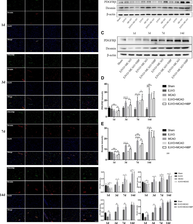
Figure 8.
Cerebral venous circulation disturbance aggravated pericytes loss, and NBP treatment could protect pericyte from severe loss in rats of MCAO accompanied by EJVO. (A) the expression of pericytes labelled by colocalisaton of PDGFRβ (green) and desmin (red) in the ischaemic area or corresponding region. (B, C) Expression of PDGFRβ and desmin detected by Western blot. (D) Quantification analysis of PDGFRβ analysed by immunofluorescence staining. (E) quantification analysis of desmin analysed by immunofluorescence staining. (F, H) Quantification analysis of PDGFRβ analysed by Western blot. (G, I) Quantification analysis of desmin analysed by Western blot. Data are presented as mean±SD, n=5 per group for immunofluorescence staining and n=3 per group for Western bolt. *P<0.05, MCAO vs sham; **p<0.01, MCAO vs sham; #p<0.05, EJVO +MCAO vs Sham; ##p<0.01, EJVO +MCAO vs sham; §p<0.05, EJVO +MCAO + NBP vs sham; §§p<0.01, EJVO +MCAO + NBP vs sham; ※p<0.05, MCAO vs EJVO; ※※p<0.01, MCAO vs EJVO; &p<0.05, EJVO +MCAO vs EJVO; &&p<0.01, EJVO +MCAO vs EJVO; ∫∫p<0.05, EJVO +MCAO vs MCAO; $p<0.05, EJVO +MCAO + NBP vs EJVO +MCAO; $$p<0.01, EJVO +MCAO + NBP vs EJVO +MCAO. Scale bar=50 µm. EJVO, external jugular vein occlusion; MACO, middle cerebral artery occlusion; NBP, Dl-3-n-butylphthalide.

