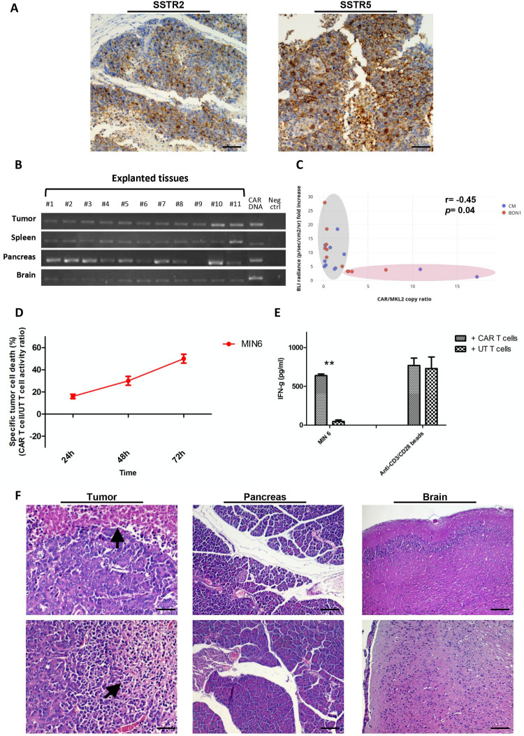
Figure 5.
Anti-SSTR CAR T cells infiltrate human xenografts and murine SSTR-expressing organs. (A) Visualization of SSTR2 (UMB1 mAb) and SSTR5 (UMB5 mAb) within tumor xenografts by IHC. Magnification: ×20. Scale bar: 100 µm. (B) Explanted CM tumor xenografts as well as SSTR-expressing organs such as murine spleen, pancreas and brain were lysed and subjected to DNA extraction. The infiltration of CAR T cells was demonstrated by PCR using primers specific for the anti-SSTR CAR sequence. The purified CAR construct DNA was used for positive control experiments. Distilled water was used in negative control experiments. (C) FAM-labeled and HEX-labeled probes against the CAR sequence and the MKL2 reference gene, respectively, were employed in ddPCR experiments to quantify the infiltration of CM (blue dots) and BON1 xenografts (red dots) by anti-SSTR CAR T cells. An inverse correlation can be observed between CAR T cell infiltration and tumor bioluminescence increase relative to baseline. (D) Anti-SSTR CAR T cells recognize and kill the MIN6 cells, a murine NET cell line expressing SSTR2/5. By in vitro BLI, CAR T cells induced cell death in the 48% of Luc+ MIN6 cells as compared with UT T cells at an E:T ratio of 1:1 after 72 hours of coculture. (E) IFN-γ release on 24 hours coculture of NET cells with CAR T cells or UT T cells at an E:T ratio of 1:1. (F) Histopathological analysis of human NET xenografts and murine pancreas and brain by H&E staining. Extensive necrosis foci could be identified within tumors (arrowhead). Representative microphotographs show the absence of necrosis or other tissue damages in the context of the murine pancreas and brain. Magnification: x20. Scale bar: 100 µm. **P<0.01. CAR, chimeric antigen receptor; ddPCR, droplet digital PCR; E:T, effector:target; IHC, immunohistochemistry; NET, neuroendocrine tumor; SSTR, somatostatin receptor; UT, untransduced.

