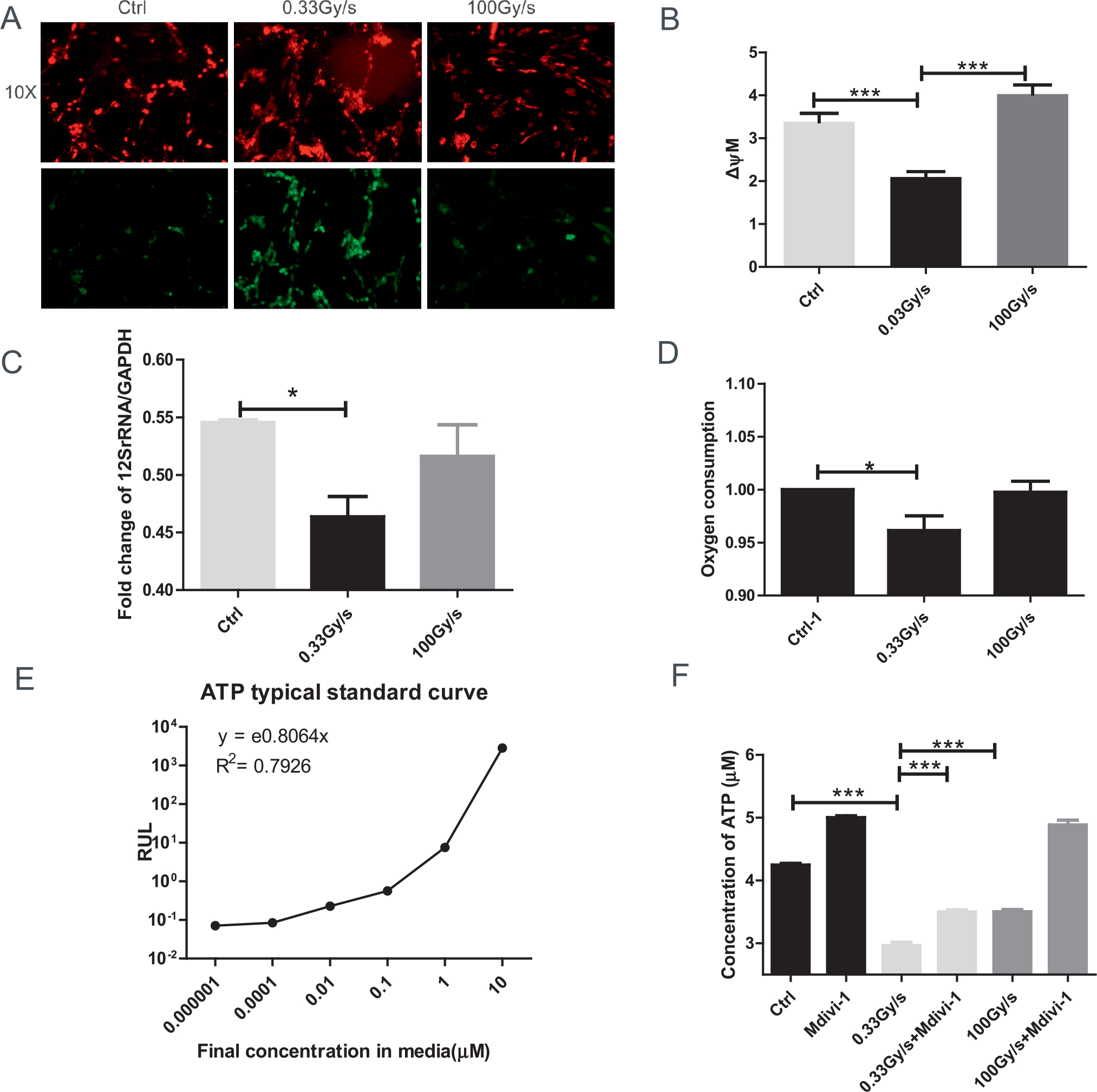
FIG. 3.

Effect of FLASH-RT on mitochondrial functions of normal cells. Panel A: Representative images of control or proton irradiated IMR90 cells delivered at 0.03 Gy/s or 100 Gy/s and stained with JC-1 (20 min at 37°C). Images are shown 200× at 590 nm (top panel) and at 530 nm (bottom panel). Panel B: Quantification of JC-1 stain (red/green fluorescence) indicative of mitochondrial membrane potential (MMP). For each sample, we acquired 5 fields of view (10×). Panel C: Fold change of mitochondrial copy number determined by real time PCR of 12S rRNA/GAPDH (24–48 h). Panel D: Oxygen consumption. Panels E and F: Total levels of cellular ATP (with typical standard curve) were analyzed immediately after irradiation. *P ≤ 0.05; ***P ≤ 0.001. For each endpoint we performed three independent experiments.
