Fig. 1.
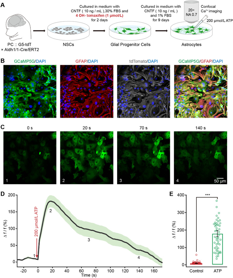
Ca2+ transients can be induced in cultured astrocytes generated from embryonic cortical NSCs. A Experimental procedure for NSC isolation, metamorphic recombination of Cre-ERT2, glial precursor cell induction, and confocal Ca2+ imaging. B Mature astrocytes (GFAP+, red) can be induced from NSCs and express both GCaMP5G (green) and tdTomato (white) following treatment with tamoxifen. Merged image indicated almost all the cells expressing GCaMP5G are GFAP positive. (C) Ca2+ imaging of cultured astrocytes derived from NSCs labeled with GCaMP5G (green) at 4 time points after addition of ATP. D Ca2+ signals evoked by 200 μM ATP in the derived astrocytes (n = 49 cells). The 4 time points shown in panel c are labeled on the trace of Ca2+ signals. E Bar graphs of astrocytic Ca2+ amplitude (Δf/f) without (control) or with ATP (n = 49 cells in each group; Control versus ATP, Z = − 6.903, P = 1.1101 E−09; ***P < 0.001, two-sided Wilcoxon signed-rank test). All data in the figure are shown as mean ± s.e.m

