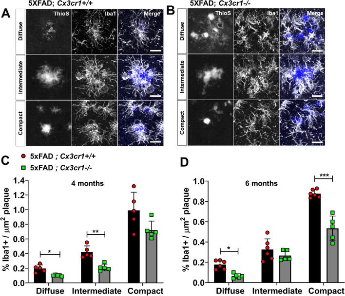
Fig. 2.
Impaired microglial plaque engagement in 4- and 6-month old 5xFAD mice deficient in Cx3cr1. Co-labeling of ThioS+ plaques and Iba1+ microglia in the cortex of 6 month-old (A) 5xFAD;Cx3cr1+/+ and (B) 5xFAD;Cx3cr1−/− mice. Images representative of cortical plaques visualized using 6 mice (3 females, 3 males) per genotype. Scale bars = 30 µm. Iba1 occupancy of the area within regions-of-interest (ROI) traced along the boundaries of diffuse, intermediate, and compact plaques calculated for plaques in the cortex of (C) 4 month-old and (D) 6 month-old 5xFAD;Cx3cr1+/+ (black bars) and 5xFAD;Cx3cr1−/− (grey bars) mice. Data in C,D represent the mean %Iba1+ area calculated within ROIs defined around cortical plaques in 4- and 6-month old 5xFAD mice with and without Cx3cr1 (n = 5–6 female and male mice of each genotype, per age). Error bars represent SEM. ~ 250–350 plaques were analyzed using multiple sections for each animal/genotype at each age. Statistical analysis done using Two-way ANOVA (pint 4 month < 0.005, p.int 6 month = 0.0002) followed by Sidak’s post hoc tests. ***p < 0.0001, **p < 0.001, *p < 0.01

