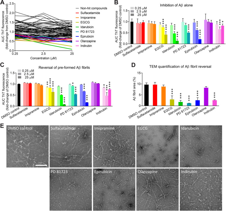
Fig. 2.
Identification of small molecule compounds that inhibit the apoE4-Aβ interaction or reverse Aβ42 fibril formation. a Dose-response experiments with 87 compounds identified in the exploratory screen that were BBB-permeable. Small molecule compounds were added at 0.25, 2.5, and 25 μM in a final concentration of 5% (v/v) DMSO. Eight hit compounds (colored lines) were identified (i.e., sulfacetamide, imipramine, EGCG, idarubicin, PD 81723, epirubicin, olanzapine, and indirubin). The data represent the mean of n = 3 wells per concentration for each compound, relative to the mean of n = 8 wells for the DMSO control. b Eight hit compounds were tested for inhibition of Aβ42 fibrillization independent of apoE4. c Eight hit compounds were tested for disaggregation of pre-formed apoE4-catalyzed Aβ42 fibrils. b, c The experiment was replicated twice, and the results were combined. The data represent the mean ± SD of n = 8 wells per concentration for each compound. *P < 0.05, **P < 0.01, and ***P < 0.001 compared to the DMSO control by one-way ANOVA. d ApoE4-catalyzed Aβ42 fibrils were treated for 30 min with DMSO or with each compound at 25 μM, except for indirubin, which was tested at 2.5 μM because it had the greatest effect in previous experiments. TEM images were acquired and analyzed for the Aβ fibril area (%). The data represent the mean ± SD of n = 3 separate TEM images for each compound. ***P < 0.001 compared to the DMSO control by one-way ANOVA. e Representative TEM images of apoE4-catalyzed Aβ42 fibrils treated with DMSO or with each hit compound. Aβ fibrils are observable by negative-stain TEM as light objects on a dark background. Scale bar = 200 nm

