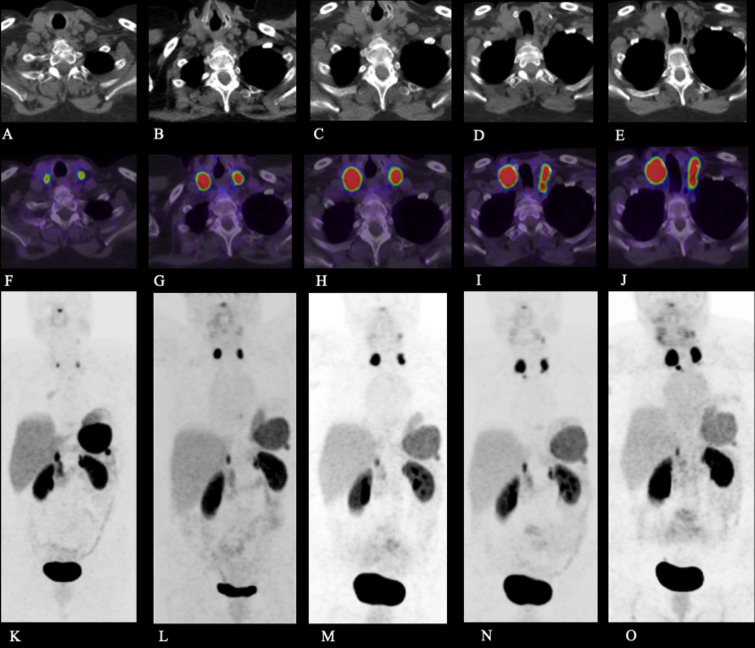
FIG. 1.
Case 1. Axial CT (A–E), fused axial [68Ga]-DOTATATE PET/CT windowed at maximum standardized uptake value (SUV) 0-15 (F–J), and [68Ga]-DOTATATE PET maximum intensity projection (MIP) image (K–O) at the following time points post–initial resection: 35 months (A, F, K), 42 months (B, G, L), 48 months (C, H, M), 54 months (D, I, N), and 58 months (E, J, O). The images demonstrate intensely DOTATATE-avid bilateral cervical lymph nodes, compatible with cervical nodal metastases.

