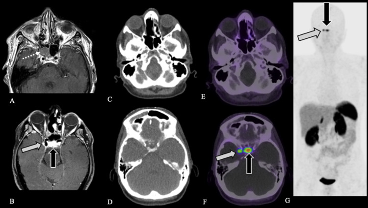
FIG. 2.
Case 3. Preoperative axial contrast-enhanced T1-weighted MRI (A) demonstrates patient’s initial lesion within the right sphenoid sinus (white dotted arrow), pathology-proven ENB. Postoperative axial contrast-enhanced fat-saturated T1-weighted MRI (B) demonstrates dural-based plaque-like enhancement along the right cavernous sinus (white arrow) and normal enhancement of the pituitary gland (black arrow). Axial CT (C and D) and fused axial [68Ga]-DOTATATE PET/CT windowed at SUV 0-15 (E and F) demonstrate expected postsurgical appearance of the sinuses without evidence of DOTATATE avidity to suggest residual or recurrent disease (C and E) as well as a focus of intense DOTATATE avidity, SUV 11.0, along the right cavernous sinus (white arrow, F), corresponding to the dura-based plaque-like enhancement (B), compatible with a meningioma. Physiological DOTATATE avidity is noted in the pituitary gland (black arrow, F). [68Ga]-DOTATATE PET MIP (G) demonstrates normal physiological uptake in the pituitary gland (black arrow) and a focus of DOTATATE avidity corresponding to the right cavernous sinus meningioma (white arrow) without evidence of recurrent or metastatic disease.

