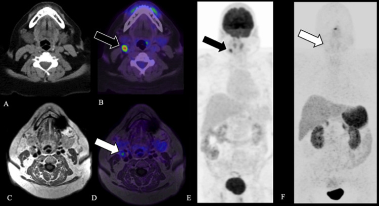
FIG. 3.
Case 4. Axial CT (A), fused axial [18F]-FDG PET windowed at SUV 0-15 (B), and [18F]-FDG PET MIP (E) demonstrate a moderately FDG-avid right level IIa lymph node, SUV 16.3 (black arrow, B, E). Axial T1-weighted MRI (C), fused axial [68Ga]-DOTATATE PET windowed at 0-15 (D), and [68Ga]-DOTATATE PET MIP (F) demonstrate a right level IIa lymph node without corresponding avidity (white arrow, D, F), favoring an inflammatory etiology. Surgical excision of the right level IIa lymph node confirmed follicular hyperplasia without evidence of malignancy.

