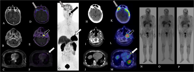
FIG. 4.
Case 5. Axial contrast-enhanced T1-weighted MRI (A–C), fused axial [68Ga]-DOTATATE PET windowed at SUV 0-15 (D–F), and [68Ga]-DOTATATE PET MIP (G) demonstrate multiple intensely DOTATATE-avid foci involving the dura (A, D, G; gray arrows), left parapharyngeal space (B, E, G; black arrows), and left anterior fourth rib (C, F, G; white arrows), compatible with metastatic ENB. After the administration of [177Lu]-DOTATATE, axial CT (H–J), fused axial [177Lu]-DOTATATE SPECT windowed at SUV 0-15 (K–M; gray, black, and white arrows), and whole-body planar images obtained at the time of each [177Lu]-DOTATATE treatment (N–P) confirm uptake of [177Lu]-DOTATATE into sites of SSTR-positive metastatic disease.

