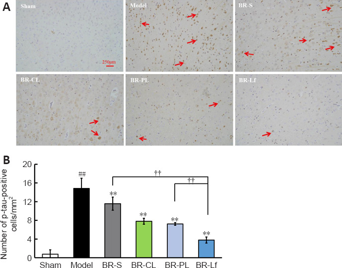
Figure 5.

Effects of different BR formulations on p-tau level in cerebral cortex of a mouse model of AD-like Aβ neurotoxicity.
(A) Immunohistochemistry images of different BR formulations on p-tau level of Ser396 (arrows) labeling in the cerebral cortex tissue of a mouse model of AD-like Aβ neurotoxicity. Of all of the groups, the BR-Lf group exhibited the optimal effects on the downregulation of hyperphosphorylated tau. Scale bars: 250 μm. (B) Quantitative analysis of p-tau-positive cells. Data are expressed as the mean ± SD (n = 4). ##P < 0.01, vs. sham group; **P < 0.01, vs. model group; ††P < 0.01 (one-way analysis of variance followed by the least significant difference post hoc test). AD: Alzheimer’s disease; Aβ: amyloid-β protein; BR: berberine hydrochloride; BR-S: BR solution; BR-CL: BR common nanoliposomes; BR-PL: BR PEGylated nanoliposomes; BR-Lf: BR PEGylated nanoliposomes with lactoferrin modified; p-Tau: Tau phosphorylation.
