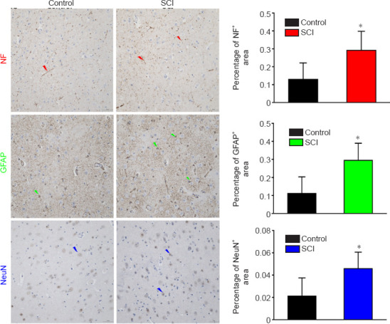
Figure 10.

Immunohistochemical staining of the PRG in a dog model of SCI at 12 weeks after injury.
The staining intensities of NF, GFAP and NeuN were all stronger, and the percentages of positively stained areas were increased significantly in the SCI group compared with those in the control group. Arrows represent typical positive expression. Arrows represent typical positive expression. Data are expressed as mean ± SD (n = 5). *P < 0.05, vs. control group (Student&s t-test). GFAP: Glial fibrillary acidic protein; NF: neurofilament heavy polypeptide; PRG: precentral gyrus; SCI: spinal cord injury.
