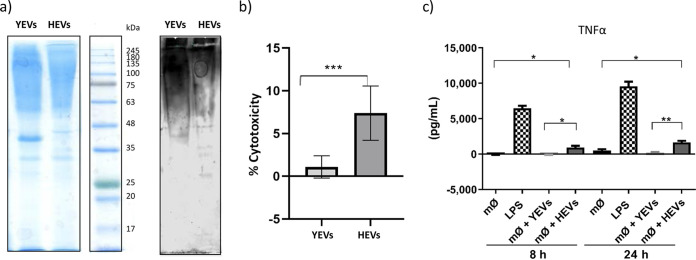
FIG 7.
Differences in the immune response against YEVs and HEVs. (a) SDS-PAGE Coomassie-blue stained gel and Western blot showing the immunoreactive patterns of YEV and HEV protein extracts to sera from patients suffering invasive candidiasis. (b) Percentage of cytotoxicity produced by either HEVs or YEVs to TPH1 macrophages. (c) TNF-α release from TPH1-activated macrophages (mØ) incubated for 8 or 24 h with 5 μg of YEVs or HEVs. Negative and positive controls (with the addition of PBS and LPS, respectively) were also evaluated. A significant change is indicated as follows: *, P < 0.05; **, P < 0.01; ***, P < 0.001 (unpaired t test). Error bars show standard deviations.

