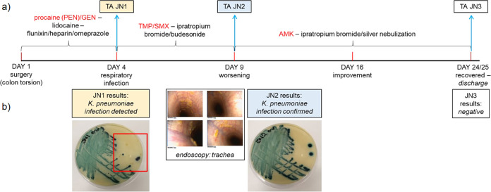
FIG 1.
Infection time course for a horse with pneumonia. (a) Severe respiratory infection developed post-surgery in a 12 -year-old mare. Antibiotic treatment was prescribed immediately and optimized over the course of 4 weeks until the infection resolved after administration of costly amikacin (AMK). Peri- and post-operative care included intravenous fluids and lidocaine by constant rate infusion, regular gastric decompressions by nasogastric intubation, penicillin (intramuscular procaine (PEN), 22 mg/kg q12H), gentamicin (intravenous GEN, 6.6 mg/kg q24H), non-steroidal anti-inflammatory drugs (intravenous Flunixin, 1.1 mg/kg q12H), enoxaparin (subcutaneous Clexane 0,5mg/kg q24H), biosponge (Di-Tri-Octahedral Smectite, q8H orally), omeprazole (gastrogard, 4 mg/kg q24H oral) and altrenogest 0.22% (Regu-Mate, 11 mL q24H oral) for the 3 days. On day 4 post-surgery, a tracheal aspirate (TA JN1) was obtained and cultured on sheep’s blood agar. PEN/GEN was substituted by oral trimethoprim-sulfamethoxazole (TMP/SMX, 30 mg/kg q12H) and ipratropium bromide and budesonide nebulization. On day 9, endoscopy was performed (b) showing increased secretions in the trachea and results of JN1 testing confirmed K. pneumoniae infection. TMP/SMX treatment was stopped, silver nebulization started, in addition to continued ipratropium bromide and budesonide therapy, and a second TA specimen (JN2) was collected. On the following day, nebulized AMK treatment was started (3.3 mg/kg q24H). Although JN2 microbiology results confirmed K. pneumoniae infection (b), the horse’s condition steadily improved until discharge on day 25, when a third TA was collected (JN3; culture-negative). Microbiological testing on CHROMagar Orientation (b) confirmed the presence of K. pneumoniae among three other species (one colony each, red square) in JN1 and alone in JN2.

