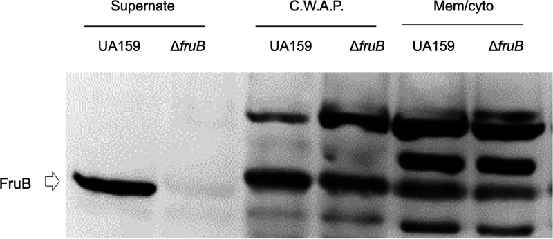
FIG 2.
Localization of FruB. S. mutans strains UA159 and △fruB were cultured in TV medium supplemented with 0.5% of inulin till mid-exponential phase. After centrifugation, the culture supernates were precipitated with trichloroacetic acid, washed and resuspended in 0.1 N NaOH. Bacterial cells were enzymatically broken up and fractionated into 2 samples, cell-wall-associated proteins (C.W.A.P) and cell membrane and cytoplasm (Mem/cyto). All 3 fractions were normalized to represent similar numbers of cells from each strain. After resolution of the protein samples on an SDS-PAGE, presence of FruB protein was visualized via Western blotting using affinity-purified anti-FruB antiserum.

