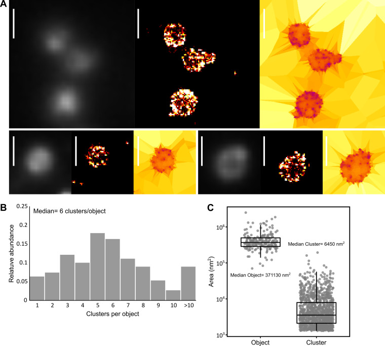
FIG 3.
dSTORM imaging of an antibody-specific protease in Mycoplasma mycoides subsp. capri. M. mycoides subsp. capri 0582-HA cells expressing an HA tag-fused variant of the serine protease MIP82 were immunolabeled and imaged by dSTORM. The tagged fusion protein is expressed from the native genomic locus and replaces the wild-type variant. The data presented here correspond to a single representative field of view (512 by 512 pixels; pixel size = 160 nm). (A) Sample images of M. mycoides subsp. capri 0582-HA cells. For each field of view, the images correspond to epifluorescence (diffraction limited) (left), superresolved reconstruction (40 nm pixel) (middle), and Tesseler segmentation (right). Scale bar = 1 μm. (B) Tesseler clustering of the fluorescence signal. For each field of view, the number of clusters per Tesseler-segmented object was computed. The bar graphs display the distribution of the numbers of clusters per object. (C) Object and cluster sizes. The dot plot presents the area (in nm2) of each object and cluster segmented by Tesseler, to which a boxplot showing the median, interquartile range, minimum, and maximum values is overlaid. The median value of each data set is indicated.

