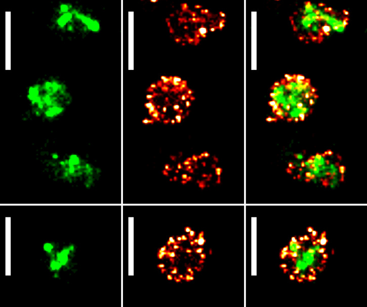
FIG 4.
PALM/dSTORM two-color imaging of Mycoplasma mycoides subsp. capri. Sample images of M. mycoides subsp. capri 0582-HA pMT85-PSynMyco-mEos3.2 cells, expressing both an HA tag-fused variant of the serine protease MIP0582 and the fluorescent protein mEos3.2. The tagged fusion protein is expressed from the native genomic locus and replaces the wild-type variant. mEos3.2 is expressed from a transposon inserted at a random site in the bacterial chromosome. For each field of view, the images correspond to a reconstructed PALM image (40-nm pixel) (left), a reconstructed dSTORM image (40-nm pixel) (middle), and an overlay of the reconstructed PALM and dSTORM images (right). Scale bar = 1 μm. All the images were sampled from the same coverslip and field of view.

