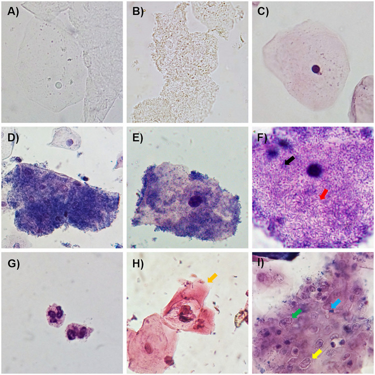
FIG 1.
Examples of the usefulness of the A-S dye solution in the diagnosis of BV. (A and B) Fresh observation of a normal squamous epithelial cell (A) and clue cells (B) in a nonstained sample. (C to E) Normal squamous epithelial cells (with normal morphology) and examples of clue cells in samples from patients with BV using the A-S dye solution. (F) Bacterial morphotypes attached to the surface of epithelial cells, including curved bacilli (red arrow) and a coccobacillus (black arrow). (G) Leukocytes (polymorphonuclear). (H) Presumptive koilocyte (associated with HPV infection) with karyomegaly and perinuclear halo (yellow arrow). (I) Group of squamous epithelial cells with inflammation-related changes such as karyomegaly (green arrow) and perinuclear halo (yellow arrow) during Candida infection (blue arrow) (1000X amplification).

