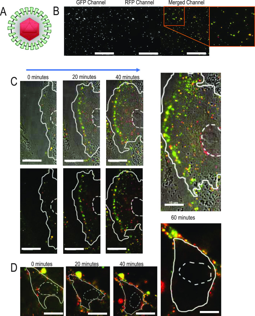
FIG 3.
Live fluorescence microscopy observes absence of nuclear localized capsids during SIE. (A) Model of PRV 137 virion containing an mRFP-VP26 fusion (red capsid) and a gM-eGFP fusion (green envelope protein). (B) Distribution of PRV 137 fluorescent assemblies from a viral supernatant preparation. Images are split into designated fluorescent channels and composite image. (C) Select images from a time course of direct infection with PRV 137 on PK15 cells. Images taken every 20 min. The top row corresponds to brightfield and fluorescent composite images, while the bottom row demonstrates only fluorescent merged images. The nucleus is outlined with dashed lines, while the cell boundary is outlined with a solid line. (D) Select images from a time course of PRV 137 during SIE. Images were taken every 20 min after application of PRV 137. Cell boundaries are as depicted above.

