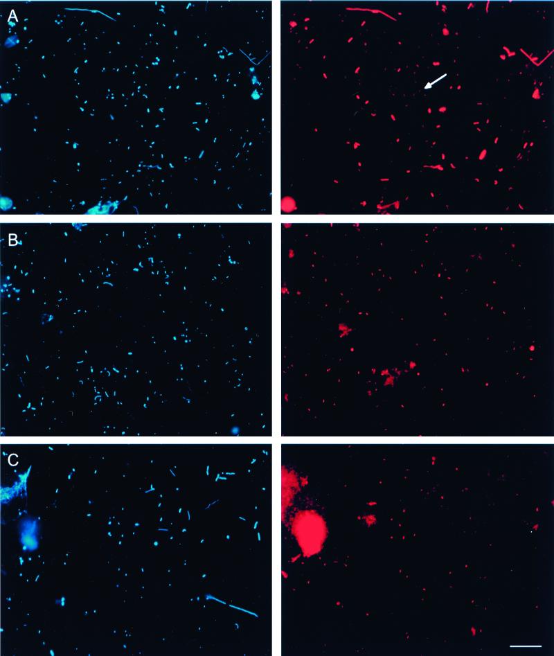
FIG. 3.
FISH of samples from Lake Gossenköllesee. DAPI (left) and epifluorescence (right) micrographs are shown for identical microscopic fields. (A) In situ hybridization with probe EUB338 labeled with Cy3. The arrow indicates a representative of a population of small, dim cells. (B) In situ hybridization with probe HGC664 labeled with Cy3. (C) In situ hybridization with probe HG1-840 labeled with Cy3. Bar, 10 μm (all panels).

