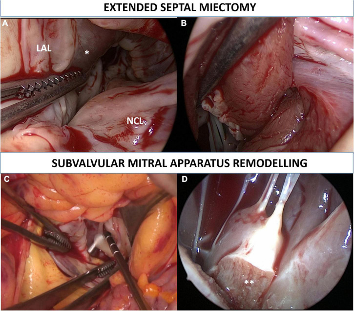
FIGURE 1.
Extended septal myectomy. Surgical view of the left ventricle outflow tract before the procedure is depicted in panel (A). Transaortic myectomy was performed in all patients starting at nadir of the right coronary sinus, and extended apically to achieve exposure of the papillary muscles bases. Muscular resection was extended toward the lateral ventricular wall up to the left trigone (B). Subvalvular mitral apparatus remodeling included (1) resection of fibrotic, thickened, and agglutinated secondary chordae tendineae from the tip of the papillary muscles to the ventricular surface of the anterior mitral leaflet (C); (2) resection of all anomalous muscular trabecula and splitting (**) of hypertrophied and thickened papillary muscles (D). LAL, left aortic leaflet; NCL, non-coronary leaflet. *Interventricular septum bulging.

