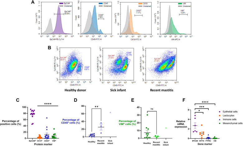
Fig. 2. Mature-stage breast milk from donors contained epithelial lactocytes and a smaller population of immune cells.
(A) Flow cytometry quantified expression of cell-specific markers: EpCAM+ (epithelial cells), CD45+ (immune cells), CK18+ (lactocytes), and VIM+ (mesenchymal cells). These data suggest that the CK18 antibody bounds poorly to lactocytes and likely underestimates true lactocyte percentages. FITC, fluorescein isothiocyanate. (B) The relative cellular composition in breast milk was affected by the health status of the mother and infant. Representative density plots are shown of EpCAM versus CD45 cell populations in milk from healthy donors (n = 10), donors with sick infants (n = 1), or donors with recent mastitis (n = 3). (C) Flow cytometry analysis of protein markers in all health statuses (n = 14) and the breakdown of (D) CD45+ immune cells and (E) VIM+ mesenchymal cells in different health statuses. (F) mRNA expression of gene markers corresponding to proteins in (C) indicates the presence of epithelial lactocytes (n = 6 to 7). Data are shown as means ± SEM. One-way analysis of variance (ANOVA) with Tukey’s multiple comparison test; *P < 0.05; **P < 0.01; ***P < 0.001; ****P < 0.0001. ns, not significant.

