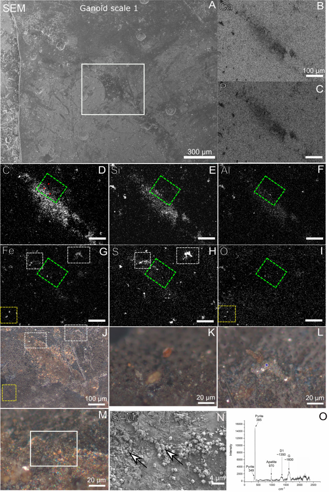
Fig 5. Chemical and mineralogical characterization of gray-brown film on Messel fossil scale by EDX and Raman.
A) SEM of gray-brown film on ganoid scale 1. Note that the ganoid scale is rotated compared with Figs 1, 2 and 4A. B-I) EDX maps of white square in A. Green square in D-I show area of ToF-SIMS analysis. Red dots in D indicate area of point spectra in S2 Table. J-M) Micrographs of ganoid scale 1. Yellow square in J indicates area of overlapping Fe and O signals and of micrograph in K. White squares in J indicate areas of Fe and S overlapping signals and of micrographs in L and M. N) SEM image of white square in M. White arrows show pyrite crystals intermixed with gray-brown film. O) Blue laser Raman spectrum obtained from blue circle in L, showing presence of pyrite in highly reflective phase infilling pore in fossil.

