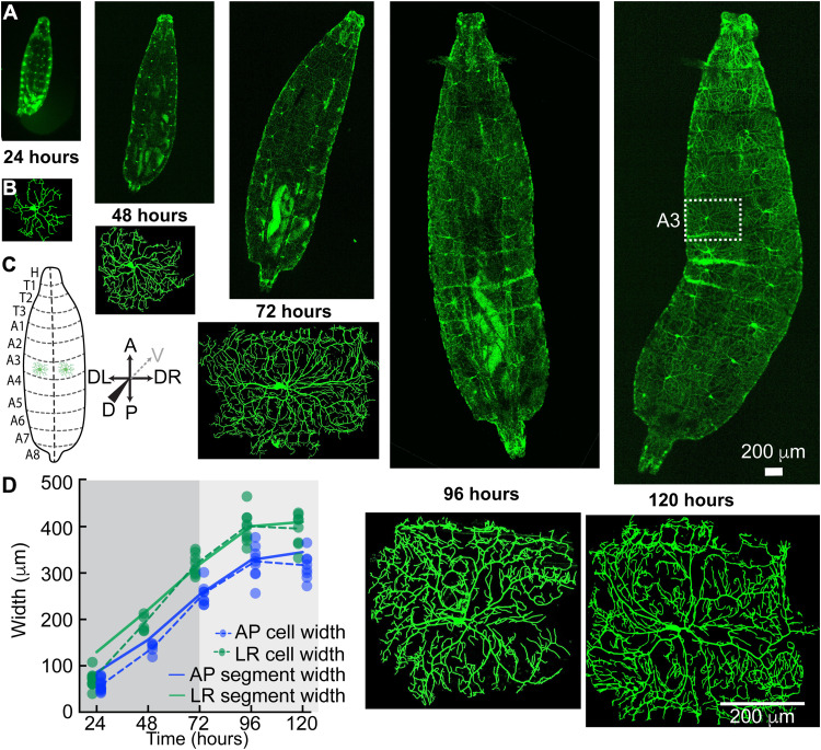
Fig. 1. Growth of larvae and class IV neurons over development.
(A) Whole-mount, living larvae imaged by spinning disk confocal microscopy at 24 to 120 hours (egg lay defined as time zero). Class IV neurons are marked with the transmembrane protein CD4 tagged with green fluorescent protein (GFP) (genotype ;;ppkCD4-tdGFP). (B) Individual class IV cells from the A3 or A4 segments. An A3 segment is outlined in (A) (120 hours). (C) Cartoon of larvae as viewed from the dorsal side. The dashed line is dorsal midline. Anterior (A) is up, and posterior (P) is down. Left (DL) and right (DR) are as viewed from the dorsal side (for the sake of simplicity, we will mention DL-DR as LR everywhere in the text and subsequent figures); the gray dashed arrow points at the ventral direction. (D) Growth of class IV arbors compared to their hemisegments. At 24 hours, the cell widths (solid circles with dashed lines through the averages) are smaller than the hemisegment widths (solid lines). In the next 24 hours, they touch the growing segment boundaries, and by 72 hours, they fill the hemisegment and then continue to grow with the hemisegment (light gray). The cell widths along each axis are defined as the sides of the rectangle that contain the same mass of branch skeleton distributed uniformly (see Materials and Methods).

