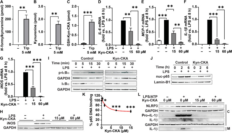
Fig. 3. Kyn-CKA is formed and attenuates NF-κB and NLRP3 signaling in macrophages.
(A to C) Kynurenine metabolites in J774a.1 8 hours after tryptophan (5 mM; n = 3). (D to G) NF-κB–dependent gene expression 8 hours after LPS (1 μg/ml) and Kyn-CKA (15 or 60 μM; n = 3). (H) Inducible nitric oxide synthase (iNOS) levels 8 hours after LPS (1 μg/ml) and Kyn-CKA. (I) Total and phospho (p)–IκBα following LPS in the presence of Kyn-CKA (0 or 30 μM). (J) Nuclear p65 (nuc-p65) in LPS-treated J774a1 versus time and Kyn-CKA (0 or 30 μM); each lane are three combined independent wells. (K) p65 (1.5 μM) DNA binding versus Kyn-CKA (n = 3 per concentration). Nonlinear fit provided for illustration purposes. (L) Inhibition of NLRP3 inflammasome activation by LPS/ATP (1 μg/ml; ATP, 2 mM) by Kyn-CKA at 8 hours. C, cellular fraction; M, media. *P < 0.05, **P < 0.01, and ***P < 0.0001 by t test (A to C) or one-way ANOVA and Tukey’s test (D to G and K).

