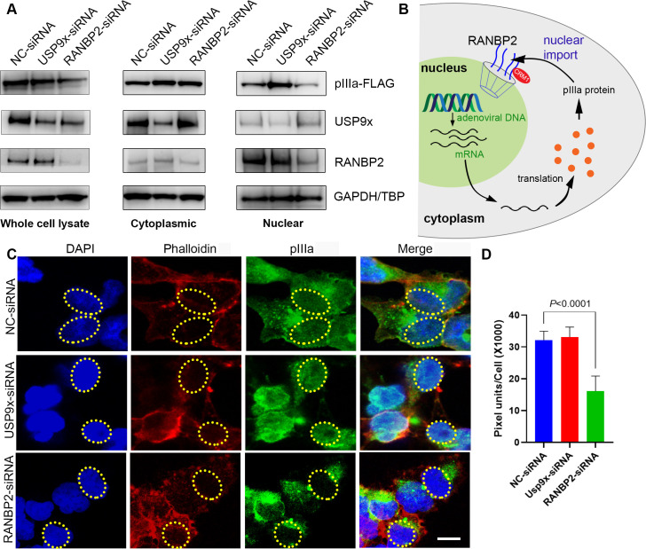
Fig 5. Human adenovirus pIIIa exploits RANBP2 nuclear import function.
(A) Cytoplasmic and nuclear fractions of stable pIIIa expressing Flp-In 293 T-Rex cells treated with USP9x or RANBP2 siRNA for 48 hours, and then induced with tetracycline (20 ng/μL), and blotted for, pIIIa-Flag, RANBP2, USP9x and GAPDH protein expression (B) Schematic of the role of pIIIa-RANBP2 interaction in the adenovirus replication cycle. (C) Following Flp-In 293 T-Rex cells pretreated with USP9x or RANBP2-siRNA were tetracycline induced (20ng/mL) for 8 hrs for pIIIa expression. FLAG-pIIIa (green), phalloidin (red), and nuclei (blue) The dotted ellipse corresponds to the nucleus area. Scale bar: 10 μm. (D) ImageJ analysis done on 30 cells per group and green fluorescence intensity was measured in the nucleus of each cell. The final data are presented as the mean ± SD of at least triplicate experiments. Statistical significance was performed with two-way ANOVA followed by Tukey multiple comparison test. Only comparisons where P<0.05 are shown.

