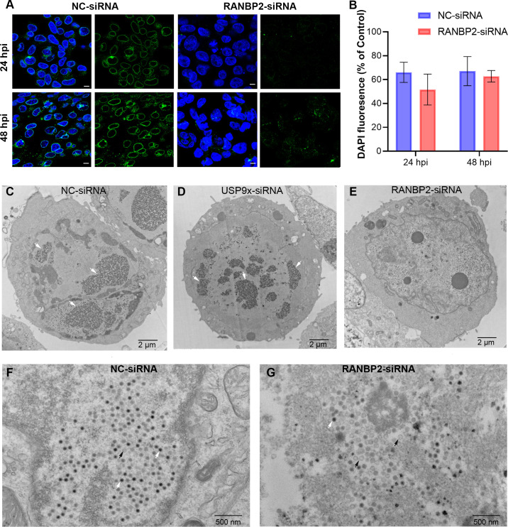
Fig 7. Human adenovirus interacts with RANBP2 for nuclear import and viral assembly.
(A) DAPI nuclear (blue) and RANBP2 (green) staining of NC-siRNA and RANBP2-siRNA treated HEK293 cells, infected with HAdV-D37 at an MOI of 0.1 for 24 and 48 hrs, respectively. Scale bar = 10 μm. (B) ImageJ quantification of DAPI fluorescence per cell, normalized to uninfected control cells (n = 100) and plotted as the percent of uninfected control fluorescence. (C) Transmission electron microscopy of NC-siRNA and (D) USP9x siRNA, and (E) RANBP2-siRNA treated HEK293 cells infected with HAdV-D37 at an MOI of 0.1 for 72 hrs. The specific MOI and time-points were chosen to establish viral replication while minimizing early cell death; HAdV-D37 infection at an MOI of ≥1 in HEK293 cells leads to significant cytopathic effect within 24 hpi. White arrows over the nuclei show large paracrystalline viral arrays. Higher magnifications are shown for NC-siRNA (F) and RANBP2-siRNA (G) treated cells. The final data in (B) are presented as the mean ± SD of triplicate experiments. Statistical significance was performed with unpaired t-test (two-tailed). No statistically significant differences were seen.

