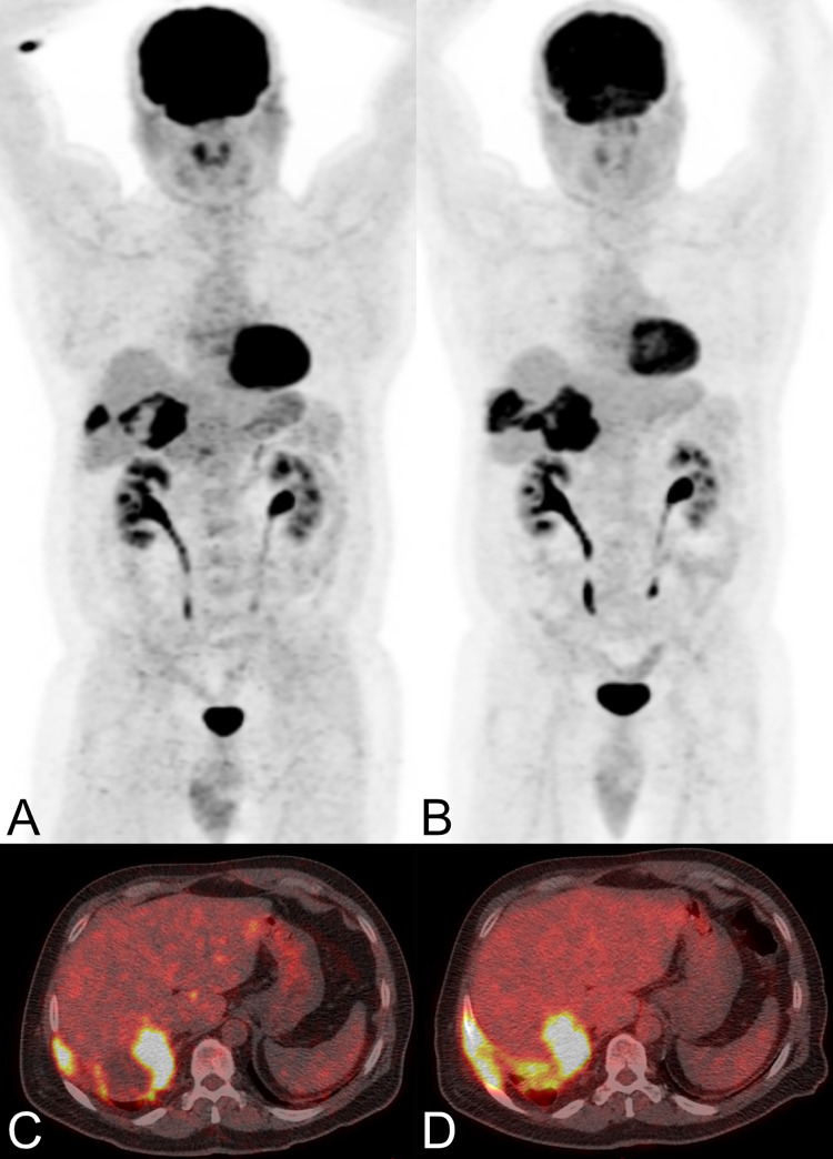
Fig 2. Two PET/CT scans performed in a male patient with a hepatic manifestation of alveolar echinococcosis involving the chest wall.
The patient (patient 09 in Table 2) was 55 years old in 2008 (maximum intensity reconstructions of PET (A) and fused PET/CT images (C)), and 58 years old in 2011 (B and D) and was treated with mebendazole from 2001 to 2011. The follow-up PET/CT 2011 (B and D) documented progression of disease with increasing involvement of the chest wall as compared to the PET/CT from 2008 (A and C) (EgHF AU increased from 34 to 35, SUVratio liver was continuously high at 4.6), and therapy was subsequently changed to albendazole. The patient died in 2017 of unknown cause.

