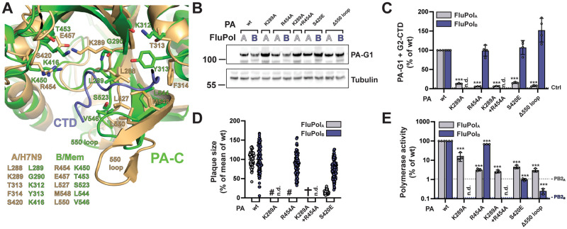
Fig 4. FluPolB and FluPolA CTD-binding mode at site 2A.
A. Superposition of CTD peptide (slate blue tube) bound at site 2A of the PA subunit of FluPolA (A/Zhejiang/DTID-ZJU01/2013(H7N9), green) with the equivalent region of FluPolB (B/Memphis/13/2003, wheat), showing similarities and differences in CTD interacting residues. See sequence alignment in Fig 1E. B-E. Protein expression (B), in vivo CTD binding (C), characterisation of recombinant IAV and IBV viruses (D) and polymerase activity (E) of CTD-binding site 2A mutants. Experiments were performed as described in Fig 3B–3E for FluPolB and FluPolA site 1AB mutations. C. ***p ≤ 0.001 (two-way ANOVA; Dunnett’s multiple comparisons test). D. (#) not measurable pinhead-sized plaque diameter; (✞) no viral rescue, (n.d.) not determined. E. ***p ≤ 0.001 (two-way ANOVA; Dunnett’s multiple comparisons test).

