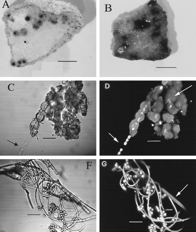
FIG. 4.
(A) Photograph of a piece of old, dried, consolidated hydrozincite with dark spots, with cyanobacterium Scytonema sp. in a dormant state. Bar, 0.5 cm. (B) The same piece of hydrozincite after a period of incubation in BG-11 medium (diluted 1:10) under dim light, clearly showing growth of Scytonema sp., which was entrapped in the hydrozincite matrix and which forms blue-green filaments. Bar, 0.5 cm. (C) In the transmission mode the Scytonema sp. clearly grows out of the hydrozincite tubing. A thick-wall heterocyst (short arrow) is clearly visible. Bar, 12, μm. (D) The same image by SCLM shows single autofluorescent cells of a Scytonema sp. Autofluorescence is due to chlorophyll a encapsulated in the sheath. Where the heterocyst occurs (short arrow), no autofluorescence appears, since the photosynthetic apparatus is degenerated. Moreover, hydrozincite emits fluorescence (long arrow). Bar, 12 μm. (F) Coculture of Scytonema sp. and the microalga Chlorella sp. The cyanobacterium shows filaments of different diameters. Bar, 12 μm. (G) The same image taken by SCLM (the sheath of the Scytonema sp. becomes empty and wide with aging and autofluorescence [arrow]). Bar, 12 μm.

