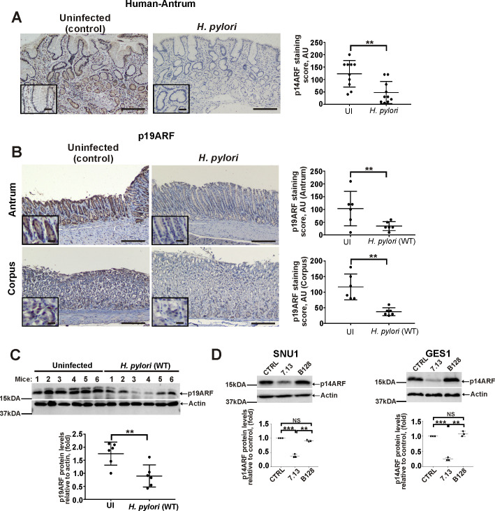
Fig 1. H. pylori infection leads to downregulation of p14ARF.
(A) Representative images showing IHC staining for p14ARF protein in antral gastric biopsies collected from H. pylori-infected and non-infected patients with active chronic gastritis. The graph panel shows IHC scores for p14ARF (n = 10/group). (B) Representative images of IHC staining for p19ARF protein in the corpus and the antrum of H. pylori-infected and uninfected mice. The graph panel shows IHC scores for p19ARF (n = 6/group). (C) The same as (B), but gastric tissue homogenates from mice were analyze for p19ARF protein expression by Western blotting. Bottom panel shows the densitometric analysis of p19ARF protein expression. (D) Analysis of p14ARF protein expression in SNU1 and GES1 cells co-cultured with H. pylori strains 7.13 or B128 for 6hrs. The graph panels represent densitometric analyses of p14ARF protein expression. Expression of p14ARF protein in uninfected cells was arbitrarily set at 1. Data in A, B, and C were analyzed using unpaired 2-tailed t-test; data in D were analyzed using 1-way ANOVA followed by Tukey’s multiple comparison test. Data are displayed as mean ± SD (n = 3). Insets in (A) and (B) show the magnified views (×40). Scale bars: 50 μm.

