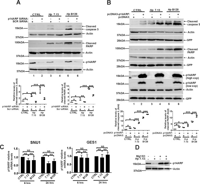
Fig 2. ARF regulates apoptosis in H. pylori infected cells.
(A) Western blot analysis of apoptosis markers, cleaved caspase 3 and PARP in SNU1 cells transfected with p14ARF siRNA or control scrambled siRNA (Scr siRNA) and then either co-cultured with H. pylori strains 7.13 or B128 for 18 hours or left uninfected. Each experiment was carried out three times (n = 3), with representative blots displayed. Bottom panels show the densitometric analyses. Protein expression levels in control cells were arbitrarily set at 1. (B) The same as (A), but AGS cells were transfected with pcDNA3-p14ARF expression plasmid or empty pcDNA3 vector. (C) qPCR analysis of p14ARF mRNA in SNU1 and GES1 cells co-cultured with H. pylori strains 7.13 or B128 for the indicated time periods. Expression of p14ARF mRNA in uninfected cells was arbitrarily set at 1. (D) SNU1 cells co-cultured with H. pylori strain 7.13 were treated with proteasome inhibitor MG132 (20μM) or DMSO (vehicle), and analyzed for p14ARF protein expression by Western blotting (n = 3). Data in (A, B and C) were analyzed using 1-way ANOVA followed by Tukey’s multiple comparison test. Data are displayed as mean ± SD.

