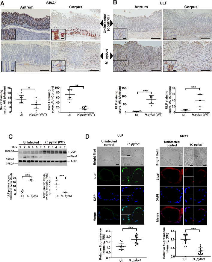
Fig 5. Analyses of ULF and SIVA1 protein expression in the murine stomach.
(A) A representative image showing IHC staining for SIVA1 protein in the corpus and antrum of H. pylori-infected and non-infected mice (n = 6/group). (B) The same as (A) but ULF protein staining is shown. (C) The same as (A and B), but Western blotting was used for analyses of ULF and SIVA1 proteins in gastric tissue homogenates. Bottom panel shows the densitometric analyses. (D) Protein expression of ULF (left panel) and SIVA1 (right panel) in gastric organoids derived from the antropyloric region of the murine stomach that were infected with H. pylori strain 7.13 in vitro or left uninfected. Panels show representative light microscopic and immunofluorescence images. Bottom panel shows quantification of ULF and SIVA1 fluorescence intensity/cell. Data were analyzed using unpaired 2-tailed t-test. Data are displayed as mean ± SD.

