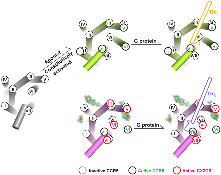
Fig. 6. The diverse active conformation of CX3CR1 and CCR5.
The helices are represented as cylinders. Cholesterol molecules are displayed in ball-stick representation with transparent spheres. The intracellular ends of transmembrane helices of inactive CCR5 (PDB ID: 4MBS), active CCR5 (PDB ID: 7F1Q), and active CX3CR1 are shown in black dashed circles, black solid circles, and red solid circles, respectively. The C termini of Gαi of CCR5-Gi and CX3CR1-Gi complexes are colored gold and slate, respectively.

