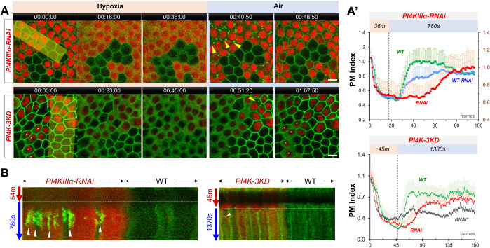
Figure 4. PI4Ks regulate the PM localization of Lgl::GFP under hypoxia and reoxygenation.
(A) Representative frames showing Lgl::GFP PM localization during hypoxia and reoxygenation. RNAi cells are labeled by RFP. Yellow arrowheads: transient Lgl::GFP puncta (only few highlighted). *: RNAi cells that failed to recover Lgl::GFP to PM. (A’) (TOP) PM localization of Lgl::GFP quantified in boundaries between wild type (WT) cells (n=23), between PI4KIIIα-RNAi (RNAi) cells (n=24) and between WT and RNAi (WT-RNAi) cells (n=24). (Figure 4—source data 1). (BOTTOM). PM localization of Lgl::GFP quantified in boundaries between wild-type (WT) cells (n=20), between PI4K-3KD (RNAi) cells (n=10) and between cells failed recovery (RNAi*) (n=10). (Figure 4—source data 2). (B) Kymograph highlights the transient Lgl::GFP puncta (arrowheads). Each kymograph was made by reslicing the movie with the maximum projection of a 150 or 250-pixel wide line (yellow bands in A). Time stamp in hr:min:sec format. Scale bars: 5 µm.

