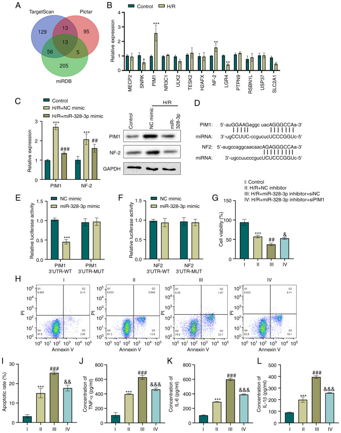
Figure 2.
miR-328-3p targets the PIM1 gene and abrogates the H/R-mediated inflammatory response in HK-2 cells. (A) Venn diagram illustrating common targeting gene candidates of miR-328-3p between the TargetScan, Pictar and miRDB databases. (B) HK-2 cells were subjected to hypoxia (12 h) and reoxygenation for 6 h. The expression levels of gene candidates were then determined using RT-qPCR. *P<0.05, **P<0.01 and ***P<0.001 vs. control group. (C) The expression levels of PIM1 and NF-2 in HK-2 transduced with NC mimic or miR-328-3p mimic and treated with H/R were examined using RT-qPCR and western blot analysis. ***P<0.001 vs. control group; ##P<0.01 and ###P<0.001 vs. H/R + NC mimic group. (D) Sequence alignment between miR-328-3p seed sequence and 3′UTR sequences of targeting genes. (E and F) Luciferase reporter gene assays were carried out with miR-328-3p mimic or NC mimic transfection in cells co-transfected with PIM1 3′UTR-WT/MUT or NF-2 3′UTR-WT/MUT. ***P<0.001 vs. NC mimic group. HK-2 cells were co-transfected with miR-328-3p inhibitor/NC inhibitor and siPIM1/siNC and then treated with H/R. (G) Cell viability was evaluated using CCK-8 assay by detecting the absorbance at 450 nm. (H and I) Cells were stabled with Annexin V or PI solutions, and cell apoptosis was detected using flow cytometry. (J-L) ELISA of the TNF-α, IL-6 and IL-1β concentrations in HK-2 cells. Data are presented as the mean ± standard deviation from three independent experiments. ***P<0.001 vs. control group; ##P<0.01 and ###P<0.001 vs. H/R + NC inhibitor group; &P<0.05, &&P<0.01 and &&&P<0.001 vs. H/R + miR-328-3p inhibitor + siNC group. PIM1, pim-1 proto-oncogene; NF-2, neurofibromin 2; RT-qPCR, reverse transcription-quantitative PCR; H/R, hypoxia/reperfusion; WT, wild-type; MUT, mutant-type; IL-1β, interleukin-1β; IL-6, interleukin-6; TNF-α, tumor necrosis factor-α.

