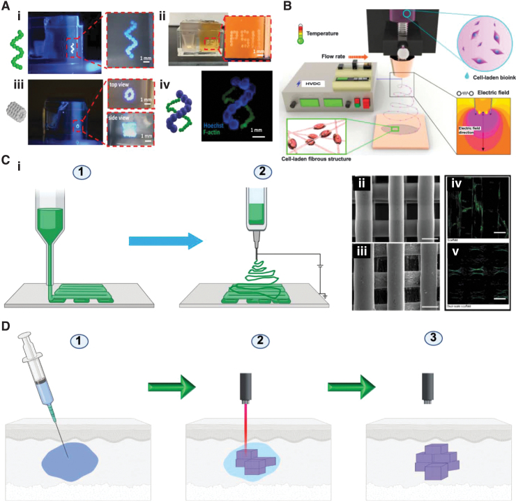
FIG. 6.
Various fabrication techniques for tissue engineering constructs. (A) Use of mesenchymal stem cell spheroids to 3D bioprint a helix shape (i), Penn State University initials (ii), 5-layer tubular structures (iii), and double helix-shape constructs (iv). One hundred fifty micrometers (F-actin) and 450 μm (Hoechst) in radius in 1.2% Carbopol yield-stress gel. Magnified zone is indicated by dashed red line. Reproduced from Ayan et al.,276 which is licensed under a Creative Commons Attribution 4.0 International License (http://creativecommons.org/licenses/by/4.0/). (B) Cell-electrospinning process with the processing parameters. Reproduced from Hong et al.,432 which is an open access article distributed under the terms and conditions of the Creative Commons Attribution (CC BY) license (http://creativecommons.org/licenses/by/4.0/). (C) Combined electrospinning and 3D printing. (i) Schematic illustration of the composite scaffold with electrospinning and 3D printing. Step (1) polymer polycaprolactone was used to 3D print the constructs and electrospinning to produce nanofibers, resulting in the formation of dual-scale scaffolds. Created with Biorender.com. Scanning electron microscope images of the scaffolds that were produced by using 3D printing (ii) and dual-scale scaffolds that were produced by using electrospinning and 3D printing (iii) (scale bar = 300 μm). Confocal laser scanning microscopy images of the scaffolds that were produced by using 3D printing (iv) and dual-scale scaffolds that were produced by using electrospinning and 3D printing (v) (scale bar = 300 μm). Reproduced from Vyas et al.,433 which is an open access article distributed under the terms of the Creative Commons CC BY license (ii–v). (D) Intravital 3D bioprinting, which is carried out by injecting a solution of the polymer into a certain tissue site to be treated in a living body. In this example, a two-photon excitation is used for the construction of a 3D object by gelating the polymer solution, and object intravital imaging is used for identification. Created with Biorender.com. Color images are available online.

