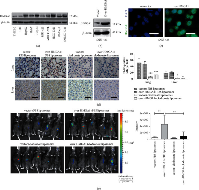
Figure 2.

Depletion of macrophage mitigates HMGA1-induced tumor growth in HCC. (a) Western blotting analysis showed the protein level of HMGA1 in eight liver cancer cell lines and two normal control cell lines (LO2 and THLE-2). (b) SUN-423 cells were transfected with ov-HMGA1 or empty vector lentivirus, and Western blotting analysis showed the protein level of HMGA1 in ov-vector and ov-HMGA1 SUN-423 cells. (c) Immunofluorescence analysis showed the protein level and distribution of HMGA1 in ov-vector and ov-HMGA1 SUN-423 cells; scale bar: 10 μm. (d) An orthotopic xenograft model was generated to determine the in vivo effect of HMGA1 in HCC; immunohistochemical analysis was performed to investigate the F4/80-positive cells in liver and lung tissues obtained from C57BL/6J mice treated with either clodronate liposomes or PBS liposomes; scale bar: 50 μm. (e) In vivo imaging analysis of tumors from the ov-vector, ov-HMGA1, ov-vector plus macrophage deletion, and ov-HMGA1 plus macrophage deletion groups (n = 5 per group). The ANOVA followed by post hoc Tukey's multiple comparison test was used for group comparisons. ∗P < 0.05; ∗∗P < 0.01; ∗∗∗P < 0.001.
