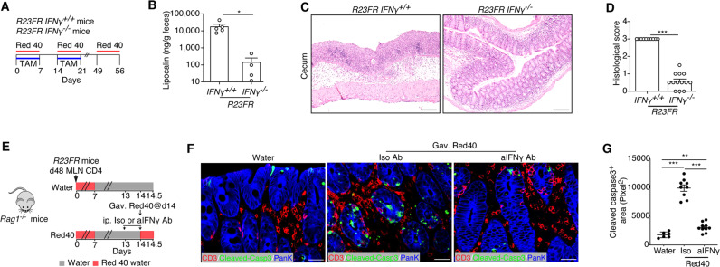
Fig. 8.
IFN-γ is required for CD4+ CTL generation and activation in vivo. A Schematic representation of the experiment to test the role of IFN-γ in CD4+ CTL generation. B Fecal lipocalin-2 levels of R23FR/Ifng-/- mice and R23FR/Ifng+/+ mice after TAM + Red 40 treatment (Day 56). n = 4–5 mice/group. C and D Representative H&E-stained sections (C) and histological scores (D) of the ceca of R23FR/Ifng−/− mice and R23FR/Ifng+/+ mice after TAM + Red 40 treatment (Day 56). n = 10–13 mice/group. Scale bars = 100 μm. E Schematic representation of the experiment to investigate the numbers of apoptotic epithelial cells after anti-IFN-γ blockade. One milligram of anti-IFN-γ or isotype antibody was administered by intraperitoneal injection prior to Red 40 gavage. F Visualization of T cells (CD3+ cells) and apoptotic (Cleaved Caspase-3+) epithelial (pan-keratin+) cells in the ceca of anti-IFN-γ-treated Red-40-gavaged adoptively transferred Rag1−/− mice. Scale bars = 25 μm. G Cleaved Caspase-3+ areas in the ceca of anti-IFN-γ and isotype antibody-treated Rag1−/− mice. n = 6–10 mice/group. Each dot represents one mouse. *p < 0.05, **p < 0.01, ***p < 0.001, by nonparametric Mann–Whitney test

