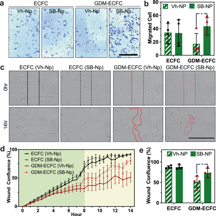
Fig. 4. Bioactive nanoparticles improve cell migration.
a Transwell migration assays were performed with normal ECFCs and GDM-ECFCs conjugated with Vh-NPs or SB-NPs. Photomicrographs depict migrated ECFCs stained with crystal violet. Scale bar is 1 mm. b The number of migrating cells after 4 h were quantified. Four biological replicates (n = 4; mean ± s.d.) were used for normal ECFCs (black data dots) and GDM-ECFCs (red data dots). SB-NPs significantly improve cell migration of GDM-ECFCs (*P = 0.039), but not normal ECFCs (P = 0.955). c Wound healing assays were performed with normal ECFCs and GDM-ECFCs treated with Vh-NPs or SB-NPs. High contrast brightfield images depict migrated ECFCs at 0 h and 14 h post wound initiation. Scale bar is 1 mm. d Kinetic wound confluence curves indicate wound closure for normal ECFCs and GDM-ECFCs treated with Vh-NPs or SB-NPs (mean ± s.d.). e Quantification of wound confluence at 8 h post wound initiation. Four biological replicates (n = 4; mean ± s.d.) were used for normal ECFCs (black data dots) and GDM-ECFCs (red data dots). SB-NPs significantly improve wound closure of GDM-ECFCs (*P = 0.048), but not normal ECFCs (P = 0.988). Statistical significance was evaluated using Student’s t-test. Significance levels were set at *P < 0.05, **P < 0.01, ***P < 0.005.

