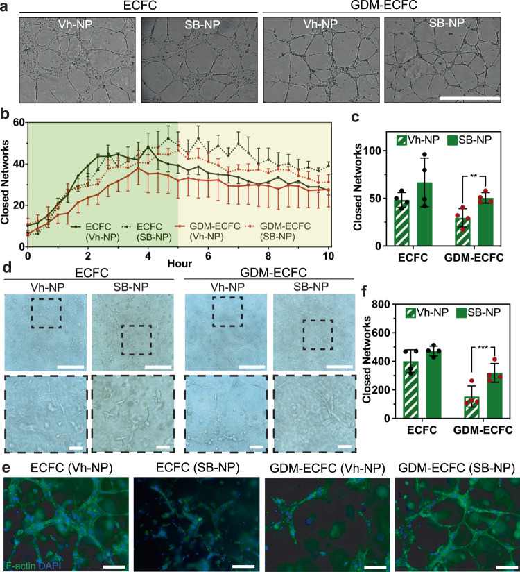
Fig. 5. Bioactive Nanoparticles Improve in vitro Vasculogenesis.
a High contrast brightfield images of ECFCs and GDM-ECFCs vascular tube formation on Matrigel 5 h post plating following treatment with Vh-NPs or SB-NPs. Scale bar is 1 mm. b Kinetic analysis of vasculogenesis (KAV) identifies closed networks formed over time. c The number of closed networks 5 h post-plating were quantified and plotted. SB-NPs significantly improve closed networks formed by GDM-ECFCs (**P = 0.010), but not by normal ECFCs (P = 0.218). Four biological replicates (n = 4; mean ± s.d.) were used for normal ECFCs (black data dots) and GDM-ECFCs (red data dots). d High contrast brightfield images of ECFCs and GDM-ECFCs forming 3D vascular networks in collagen at 48 h post encapsulation following conjugation with Vh-NPs or SB-NPs. High magnification images of the dashed areas depict the vascular tube networks. Scale bars are 100 µm. e Fluorescent images of ECFCs and GDM-ECFCs forming 3D vascular networks were stained for F-actin (green) and nuclei (blue). Scale bars are 50 µm. f The number of closed networks 48 h post encapsulation were quantified and plotted. Four biological replicates (n = 4; mean ± s.d.) were used for normal ECFCs (black data dots) and GDM-ECFCs (red data dots). SB-NPs significantly improve closed networks formed by GDM-ECFCs (***P = 0.0031), but not by normal ECFCs (P = 0.051). Statistical significance was evaluated using Student’s t-test. Significance levels were set at *P < 0.05, **P < 0.01, ***P < 0.005.

