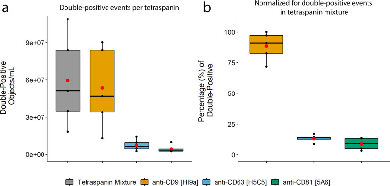
Fig. 6. Tetraspanin distribution within 5 PPP samples.
All samples were stained with CFDA-SE and an anti-tetraspanin mixture or one of the anti-tetraspanin antibodies at a concentration equal to that used in the mixture. a Tetraspanin distribution determined using anti-CD9 [HI9a], anti-CD63 [H5C5] and anti-CD81 [5A6], and (b) their relative frequencies of double-positive events compared to that obtained with the anti-tetraspanin mixture. Results shown represent events (double-positive objects/mL) obtained with each of these staining combinations and are colored as follows: gray boxes – anti-tetraspanin mixture, orange boxes – anti-CD9, blue boxes – anti-CD63, green boxes – anti-CD81. Red dots: means of sample spread. Black dots, individual PPP samples.

