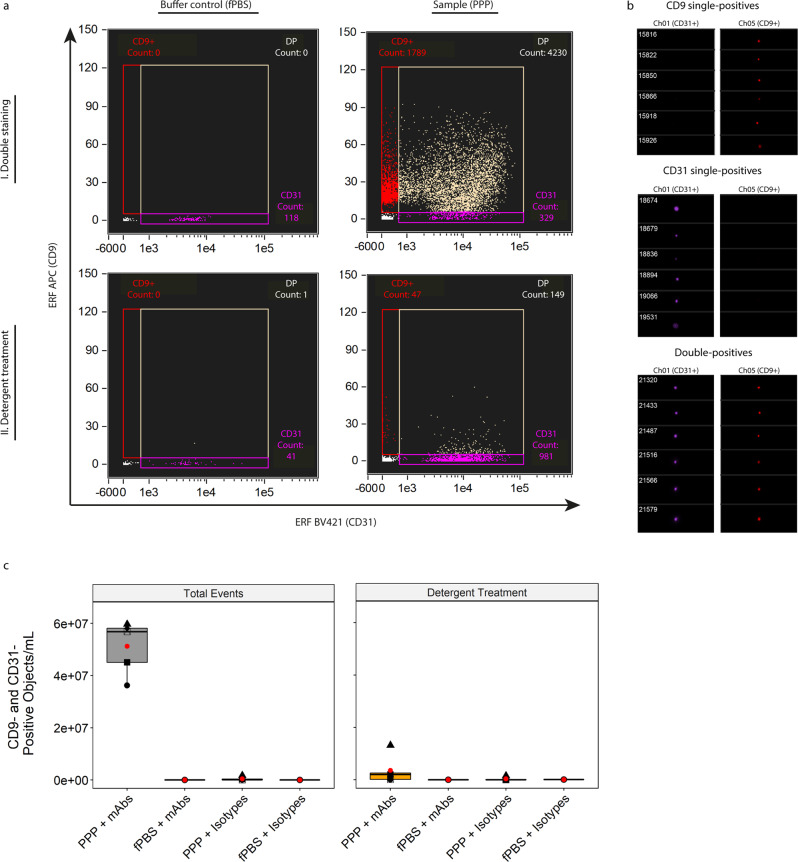
Fig. 7. Identification of single EVs on the basis of vesicular surface markers.
a Representative, fluorescence calibrated data obtained for buffer control (fPBS, left column) and PPP (right column) samples stained with anti-CD31-BV421 and anti-CD9-APC mAbs. Detergent treatment was performed by incubating the samples for 30 minutes with 20 µL 10% (v/v) TritonX-100 stock solution. Red gate: Single-positive CD9 events, purple gate: single-positive CD31 events, tan gate: double-positive events. I, double staining and II, double staining after detergent treatment. b Visual interrogation of the gated populations in the representative PPP sample. c Quantification of double-positive fluorescent events in 5 PPP samples and fPBS, stained with mAbs or isotypes, before and after detergent treatment. Approximately 93% of double-positive events in PPP stained with mAbs represent PPP-derived single EVs that were detected well above the fluorescent background. Red dots: means of sample spread. Symbols: individual PPP samples.

