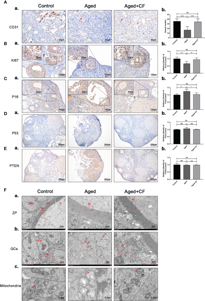
Figure 3.
CEFFE promotes angiogenesis in the medulla, promotes proliferation, and reduces senescence in ovarian granulosa cells. (A) IHC staining of the angiogenesis marker CD31 of ovarian sections (a) and statistical analysis (b) (n=4), the red arrows indicate blood vessels. Scale bar=50 μm. (B) IHC staining of the proliferation marker Ki67 of ovarian sections (a) and statistical analysis (b) (n=5-7), scale bar=100 μm. There are partial enlarged images of granulosa cells in the upper left corner of figure (a), scale bar=50 μm. (C) IHC staining of the senescence marker P16 of ovarian sections (a) and statistical analysis (b) (n=4), scale bar=100 μm. There are partial enlarged images of granulosa cells in the upper left corner of figure (a), scale bar=50 μm. (D) IHC staining of the suppressive gene PTEN of ovarian sections (a) and statistical analysis (b) (n=4), scale bar=200 μm. (E) IHC staining of the oncogenic gene P53 of ovarian sections (a) and statistical analysis (b) (n=4), scale bar=200 μm. (F) Effects of CEFFE treatment on the ultrastructural changes of zona pellucida (a), granulosa cells (b), and mitochondria (c) were observed via TEM. Scale bar=2 μm or 0.5 μm. O, Oocyte; GCs, granulosa cells; ZP, Zona pellucida; Mi, microvilli; LD, Liquid droplet; N, Nucleus; M, mitochondria. Data are represented as the mean ± SD, ns: P > 0.05, * P < 0.05, ** P < 0.01, *** P < 0.001, and **** P < 0.0001.

