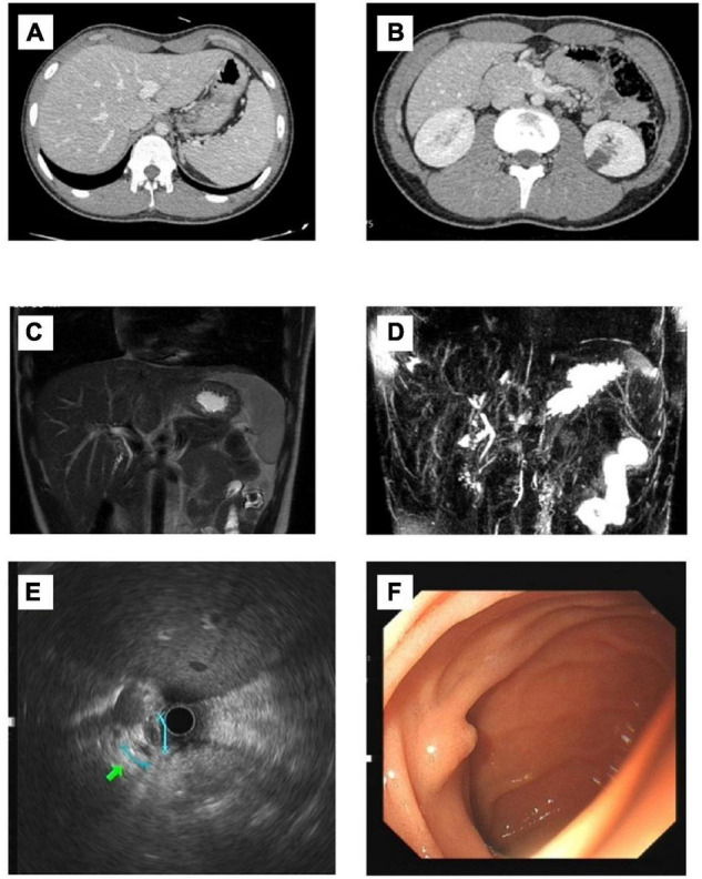
FIGURE 1.

Imaging findings of the patient. (A,B) CT: No pathological findings were found. No dilatation of intrahepatic and external bile ducts was noted. There was no thickening or enhancement of extrahepatic bile ducts. The gallbladder is collapsed without bile filling; (C,D) MRCP: Possible stenosis at the beginning of the common hepatic duct. (E) Endoscopic ultrasonography: The extrahepatic bile duct was fine without dilation, and no definite obstruction was observed. (F) Gastroscopy: The size and morphology of the duodenal papilla were normal.
