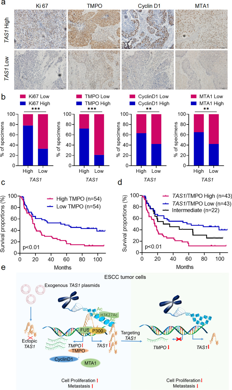
Fig. 7. Clinical relevance of the TAS1/TMPO axis in ESCC.
a Representative images of immunohistochemical staining for Ki67, TMPO, CyclinD1 and MTA1 in tissues from patients with ESCC exhibiting low or high TAS1 expression. Scale bar, 100 µm. b Percentage of specimens with low or high Ki67, TMPO, CyclinD1 and MTA1 expression in the low and high TAS1 expression groups (SYSUCC, n = 108, chi-square test, two-sided). c Kaplan–Meier analysis of OS for patients with ESCC (SYSUCC) exhibiting low (n = 54) or high (n = 54) TMPO expression (log-rank test, two-sided). d Kaplan–Meier analysis of OS for patients with ESCC (SYSUCC) exhibiting low (low expression of both TAS1 and TMPO, n = 43), high (high expression of both TAS1 and TMPO, n = 43) or intermediate (n = 22) TAS1/TMPO expression (log-rank test, two-sided). e Graphical abstract showing that the lncRNA TAS1 activates TMPO transcription in cis by recruiting FUS and p300 to modulate H3K27ac modification in the promoter region and that targeting TAS1 attenuates ESCC progression. The data are presented as the mean ± S.D. values. *P < 0.05; **P < 0.01; ***P < 0.001; ns, not significant.

