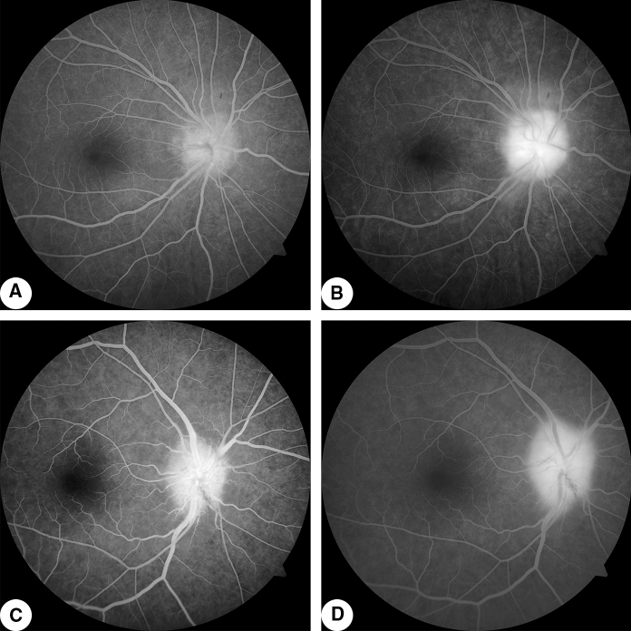
Fig. 1.
Fluorescein fundus angiogram A Case 1: early-stage right eye angiogram (0:00:29.4) with hyperfluorescence of dilated capillaries on the surface of the optic papilla; B Case 1: late angiogram (0:05:11.2) with hyperfluorescence of the whole optic papilla. C Case 2: early right eye angiogram (0:01:32.3) with hyperfluorescence of the optic papilla; D Case 2: late angiogram (0:08:25.5) with markedly enhanced fluorescence of the optic papilla

