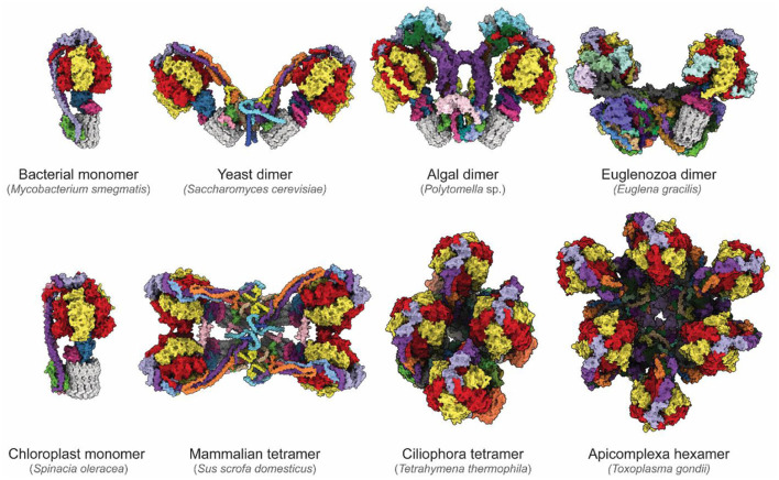
Figure 4.
Diversity of ATP synthases. Atomic models of ATP synthases from diverse species. Homologs of subunits α, β, γ, δ, ε, and c are colored red, yellow, blue, pink, purple, and gray, respectively. From the left to right, top to bottom: Mycobacterium smegmatis (PDB: 7JG5) (Guo et al., 2021), Saccharomyces cerevisiae dimer (PDB: 6B8H) (Guo et al., 2017), Polymotella sp. dimer (PDB: 6RD4) (Murphy et al., 2019), Euglena gracilis dimer (PDB: 6TDU) (Mühleip et al., 2019), Spinacia oleracea (PDB: 6FKF) (Hahn et al., 2018), Sus scofa domesticus tetramer (PDB: 6J5K, 6ZNA) (Gu et al., 2019; Spikes et al., 2020), Tetrahymena thermophila tetramer (PDB: 6YNZ) (Flygaard et al., 2020), and Toxoplasma gondii hexamer (PDB: 6TML) (Mühleip et al., 2021).

