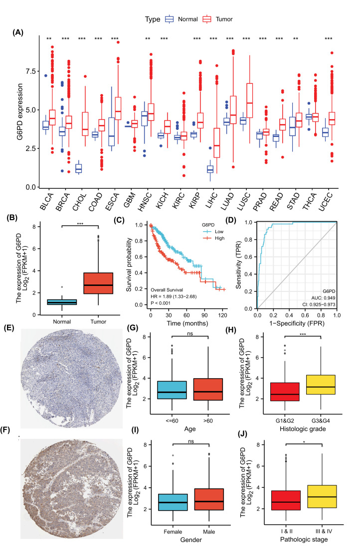
Figure 2. Expression and prognostic value of G6PD.
(A) Pan-cancer analysis, G6PD is expressed in different human tumours. (B) G6PD is expressed differently in HCC tumour tissues and normal tissues. (C) Kaplan–Meier survival curves were used to analyze the prognostic value of G6PD in HCC. (D) ROC analysis of the G6PD signatures in HCC. (E) Immunohistochemical images. The protein expression of G6PD in normal liver tissue was derived from HPA. (F) Immunohistochemical images. The protein expression of G6PD in HCC was derived from HPA. (G–J) The expression of G6PD in age, gender, histologic grade and pathological stage, respectively. ns: no sense; *: P<0.05; **: P<0.01; ***: P<0.001.

