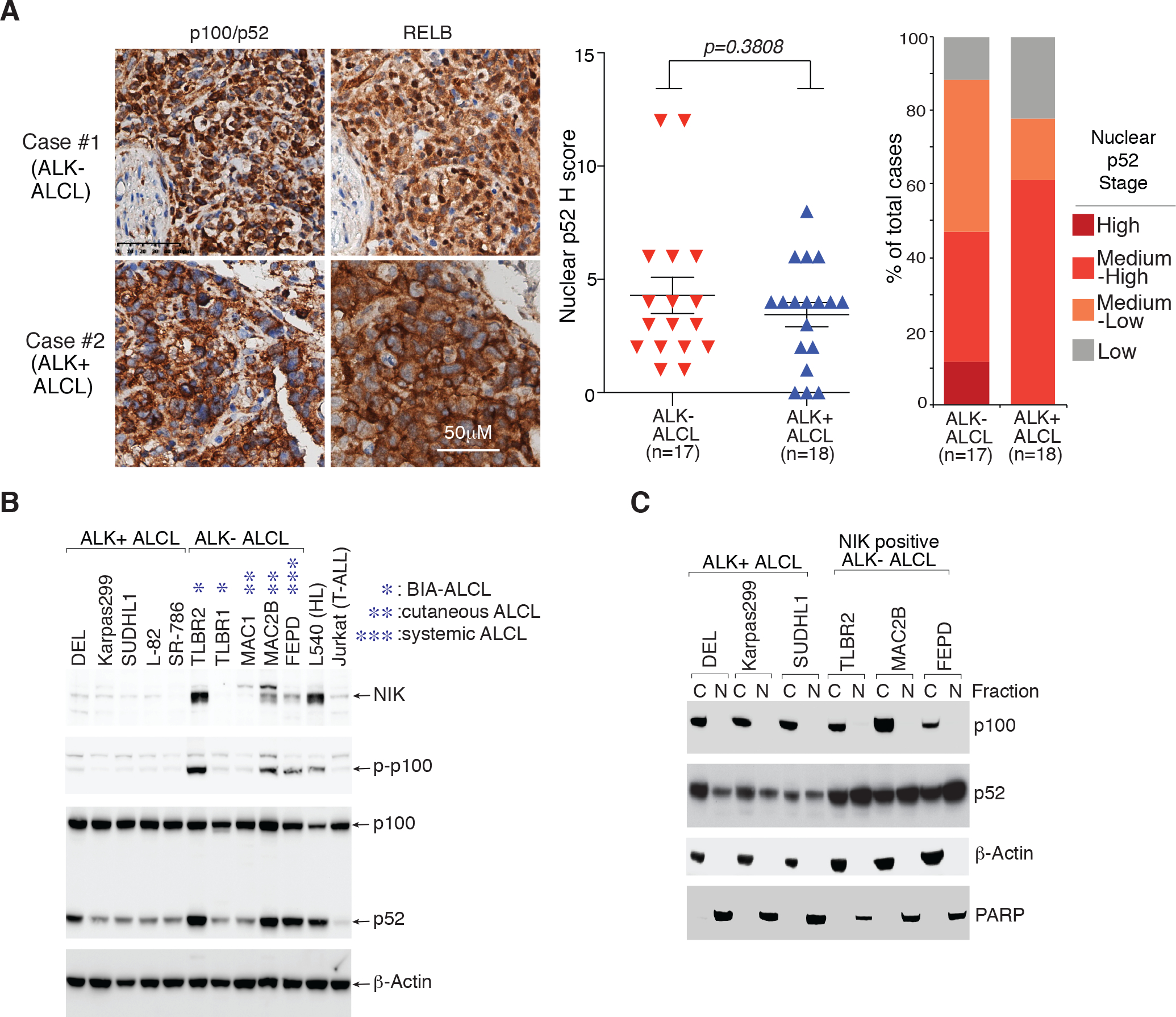
Figure 1: Expression of p100/p52 in ALCL primary samples and cell lines.

A. Expression of p100/p52 and RELB in ALCL primary cases by immunohistochemistry (IHC). left: Immunohistochemical p100/p52 and RELB staining are shown in two cases. Section of lymph nodes were examined microscopically using a 400X magnification. middle: Nuclear p52 IHC score (detailed at Supplementary Table 1) of the 17 ALK− ALCL and 18 ALK+ ALCL cases examined. p value between ALK− and ALK+ groups is indicated. right: Nuclear p52 stages in primary ALCL cases. IHC score (H Score): <2 (low); >=2, <4 (medium-low); >=4, <9 (medium-low); >=9(high). B. Lysates of indicated cell lines were analyzed by immunoblotting for the indicated proteins. C. Nuclear and cytosol fractions of indicated cell lines were analyzed by immunoblotting for the indicated proteins.
